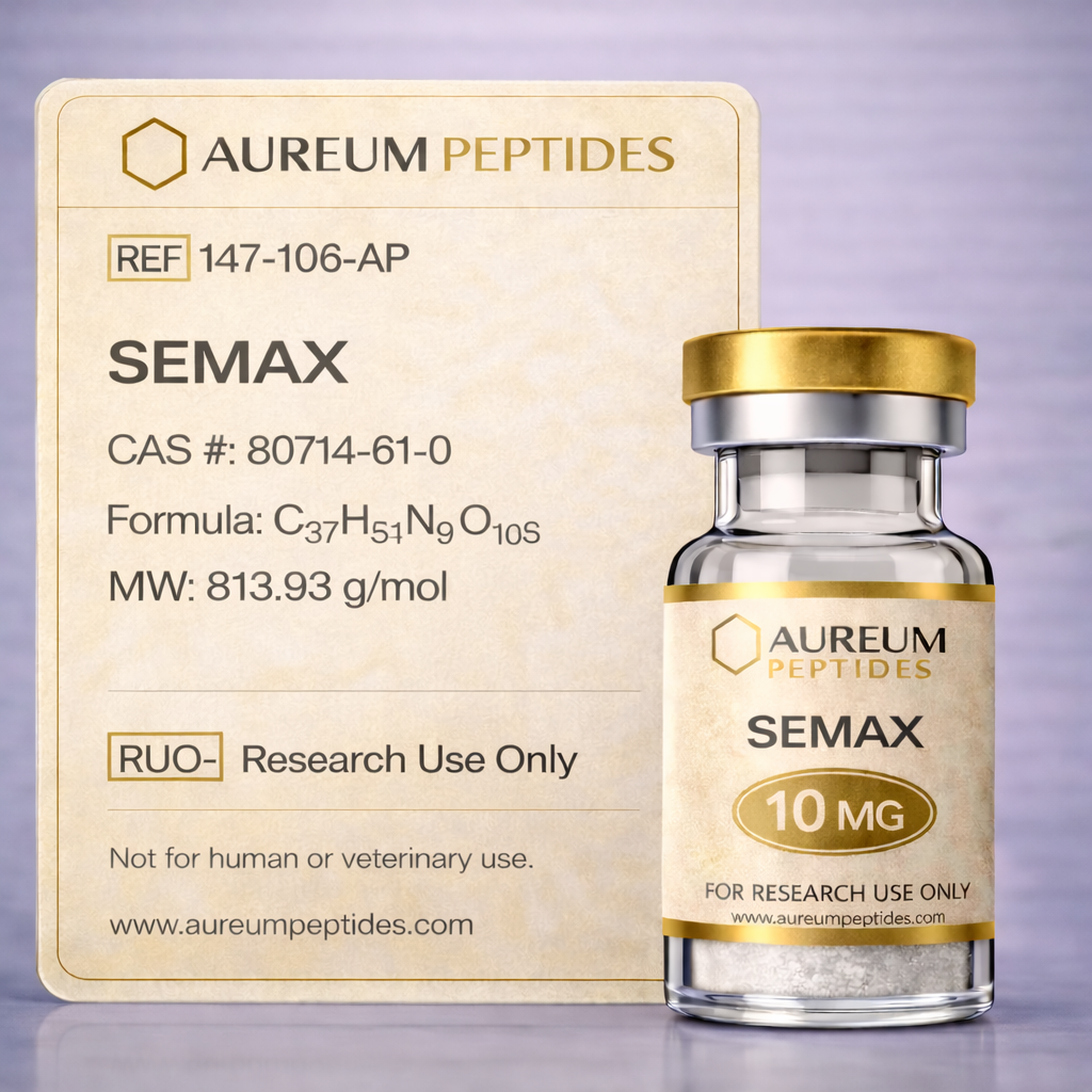
Semax
ACTH(4-10)-Pro-Gly-Pro, Semax peptide, Met-Glu-His-Phe-Pro-Gly-Pro, MEHFPGP, ACTH 4-7-PGP, Heptapeptide Semax
Quality & Transparency
Every batch tested. Every result published. No exceptions.
Semax is a synthetic heptapeptide derived from the adrenocorticotropic hormone (ACTH) fragment ACTH(4-10), with the addition of a C-terminal Pro-Gly-Pro tripeptide extension. Originally developed at the Institute of Molecular Genetics of the Russian Academy of Sciences, this neuropeptide analog has become a significant tool in preclinical neuroscience research. Semax is classified as a regulatory peptide with demonstrated stability in biological systems and blood-brain barrier penetration capability. The compound has generated substantial research interest for its modulatory effects on neurotransmitter systems, particularly dopaminergic and serotonergic pathways. Primary research applications include investigations into neuroprotective mechanisms, cognitive function modeling, neuroplasticity studies, and cerebrovascular research. In laboratory settings, Semax serves as a valuable probe for examining BDNF (brain-derived neurotrophic factor) expression, oxidative stress responses, and neuroinflammatory processes. Its unique structural characteristics and receptor interaction profile make it particularly relevant for researchers studying peptide-mediated neuromodulation and developing novel experimental paradigms in neuropharmacology and molecular neurobiology research.
Semax possesses the amino acid sequence Met-Glu-His-Phe-Pro-Gly-Pro, representing a heptapeptide with a molecular formula of C37H51N9O10S and an approximate molecular weight of 813.9 Da. The structural foundation derives from the ACTH(4-7) fragment (Met-Glu-His-Phe), which is extended by a stabilizing Pro-Gly-Pro tripeptide sequence at the C-terminus. This modification significantly enhances proteolytic resistance compared to native ACTH fragments, extending the compound's half-life in biological matrices. The presence of proline residues introduces conformational constraints that contribute to structural stability and resistance to peptidase degradation. Biochemical studies indicate that Semax does not directly bind to classical ACTH receptors (melanocortin receptors), but instead exhibits modulatory activity through interactions with neurotrophic factor systems and neurotransmitter receptors. The peptide demonstrates favorable stability in cerebrospinal fluid and plasma when reconstituted appropriately. Its amphipathic character, conferred by the presence of both hydrophobic (Met, Phe, Pro) and charged (Glu, His) residues, facilitates membrane interactions and cellular uptake in experimental models, making it suitable for diverse in vitro and in vivo research applications.
Semax has established utility across multiple domains of preclinical neuroscience research, serving as an experimental tool for investigating peptide-mediated neuromodulation and neuroprotective mechanisms. In laboratory research, this heptapeptide enables investigators to examine complex neurobiological processes at molecular, cellular, and systems levels. Preclinical studies suggest that Semax modulates multiple neurotransmitter systems and neurotrophic factor expression, providing researchers with a multifaceted probe for mechanistic investigations.
- Neurotrophic factor expression studies, particularly BDNF and NGF upregulation in neuronal cell cultures and animal models
- Dopaminergic and serotonergic system modulation research in rodent behavioral paradigms and neurochemical assays
- Oxidative stress response investigations using cellular models of excitotoxicity and mitochondrial dysfunction
- Cerebral ischemia modeling in experimental stroke research, examining penumbra preservation and inflammatory cascades
- Cognitive function and memory consolidation studies in spatial learning tasks and fear conditioning protocols
- Neuroplasticity research involving long-term potentiation, dendritic spine morphology, and synaptic protein expression
- Blood-brain barrier transport mechanism studies and peptide pharmacokinetic profiling in CNS penetration research
At the molecular level, in vitro and in vivo models indicate that Semax influences multiple signaling cascades relevant to neuronal survival and function. Research observations demonstrate that this peptide modulates the expression of neurotrophic factors, particularly brain-derived neurotrophic factor (BDNF) and nerve growth factor (NGF), through mechanisms involving transcriptional regulation. Preclinical studies suggest that Semax enhances BDNF mRNA expression in hippocampal neurons, with downstream activation of the TrkB receptor pathway and subsequent engagement of PI3K/Akt and MAPK/ERK signaling cascades. These pathways are critically involved in neuronal survival, synaptic plasticity, and dendritic arborization in experimental models.
Laboratory research has also identified Semax-mediated modulation of monoaminergic neurotransmitter systems. In rodent brain tissue analyses, the peptide has been associated with alterations in dopamine and serotonin metabolism, potentially through effects on enzymatic activity of monoamine oxidase and catechol-O-methyltransferase. Furthermore, preclinical investigations indicate that Semax influences the expression and activity of enkephalin-degrading enzymes, suggesting interactions with endogenous opioid peptide systems. Gene expression profiling studies in animal models have revealed upregulation of genes involved in antioxidant defense, including glutathione peroxidase and superoxide dismutase, indicating potential modulation of Nrf2-ARE (nuclear factor erythroid 2-related factor 2-antioxidant response element) pathway components.
Additional research observations point to anti-inflammatory pathway modulation, with in vivo studies demonstrating reduced expression of pro-inflammatory cytokines including TNF-α, IL-1β, and IL-6 in experimental neuroinflammation models. Semax has been shown in preclinical research to influence NF-κB signaling, a master regulator of inflammatory gene transcription. The peptide's effects on hypoxia-inducible factor-1α (HIF-1α) stabilization have also been documented in ischemic brain tissue models, suggesting involvement in adaptive responses to oxygen-glucose deprivation in experimental settings.
Extensive preclinical investigations have characterized Semax's molecular and cellular effects across diverse experimental paradigms. In rodent models of cerebral ischemia, including middle cerebral artery occlusion (MCAO) protocols, laboratory studies have documented reduced infarct volumes, decreased neuronal apoptosis markers (caspase-3 activation, TUNEL-positive cells), and preserved mitochondrial membrane potential in penumbral regions. These observations correlate with maintained expression of antiapoptotic proteins such as Bcl-2 and reduced cytochrome c release in neuronal cell culture models subjected to oxygen-glucose deprivation. Electrophysiological recordings in hippocampal slice preparations have demonstrated that Semax application enhances long-term potentiation (LTP) magnitude and duration, with associated increases in postsynaptic density protein-95 (PSD-95) and synapsin-I expression, suggesting modulation of synaptic structural proteins.
Behavioral neuroscience research utilizing Morris water maze, novel object recognition, and passive avoidance paradigms in rodent models has consistently shown performance enhancements in Semax-treated groups compared to vehicle controls, with corresponding neurochemical analyses revealing elevated hippocampal BDNF levels and increased dendritic spine density in CA1 pyramidal neurons. In experimental models of neurotoxicity induced by agents such as 6-hydroxydopamine or 1-methyl-4-phenyl-1,2,3,6-tetrahydropyridine (MPTP), preclinical studies have documented preservation of dopaminergic neurons in substantia nigra, maintained striatal dopamine concentrations, and reduced microglial activation markers (Iba-1, CD11b expression) in Semax-treated animals.
Molecular analyses in these research models have revealed modulation of immediate early genes including c-Fos and Arc/Arg3.1, which are implicated in activity-dependent neuronal responses and memory consolidation processes. Proteomics studies of brain tissue from Semax-treated experimental animals have identified altered expression profiles of proteins involved in energy metabolism, cytoskeletal organization, and synaptic vesicle cycling. Neurochemical quantification studies using high-performance liquid chromatography (HPLC) have documented increased norepinephrine and serotonin turnover ratios in cortical and hippocampal regions, providing mechanistic context for observed effects in preclinical behavioral assays.
Semax is supplied as a sterile, lyophilized powder for reconstitution in appropriate research-grade solvents. Each batch undergoes rigorous analytical verification to ensure consistency and quality for laboratory applications. High-performance liquid chromatography (HPLC) analysis confirms peptide purity ≥98%, with chromatographic profiles documenting the absence of significant degradation products or synthesis-related impurities. Molecular identity is verified through matrix-assisted laser desorption/ionization time-of-flight (MALDI-TOF) mass spectrometry, confirming the expected molecular weight and sequence integrity. Endotoxin testing is performed using Limulus Amebocyte Lysate (LAL) assay to ensure levels remain below specified thresholds appropriate for in vitro and in vivo research use. A Certificate of Analysis (COA) accompanies each product lot, documenting all analytical test results and providing researchers with full traceability. These quality control measures support experimental reproducibility and consistency across laboratory workflows, enabling reliable research outcomes in peptide neuroscience investigations.
- 1Ashmarin IP et al., Neuroscience and Behavioral Physiology, 1997 27(4):428-435. PubMed
- 2Gusev EI et al., Zhurnal Nevrologii i Psikhiatrii imeni S.S. Korsakova, 1997 97(6):26-34. PubMed
- 3Medvedeva EV et al., Journal of Molecular Neuroscience, 2014 52(4):538-545. PubMed
- 4Shadrina MI et al., Molecular Biology, 2010 44(6):965-971. PubMed
- 5Levitskaya NG et al., Journal of Neurochemistry, 2004 89(3):520-531. PubMed
- 6Volodina MA et al., Russian Journal of Bioorganic Chemistry, 2015 41(5):515-520. PubMed
- 7Colaço CS et al., Behavioural Brain Research, 2016 315:67-78. PubMed
- 8Alekhina TA et al., Bulletin of Experimental Biology and Medicine, 2007 143(3):318-321. PubMed
- 9Agapova TY et al., Journal of Peptide Science, 2008 14(3):317-322. PubMed
- 10Inozemtsev AN et al., Pathophysiology, 2008 15(2):149-152. PubMed
- 11Storozheva ZI et al., Bulletin of Experimental Biology and Medicine, 2008 145(5):569-571. PubMed
- 12Makarov SV et al., International Journal of Molecular Sciences, 2015 16(3):4333-4349. PubMed
Advancing Research, One Peptide at a Time
Premium quality. Rigorous testing. Trusted by researchers worldwide.

Third-party testing data will be displayed here once available.
Temperature
Lyophilized: -20°C
Reconstituted: 2-8°C (30 days)
Shelf Life
24 months lyophilized
30 days reconstituted
Handling
Avoid freeze-thaw cycles.
Use bacteriostatic water for reconstitution.
Frequently Asked Questions
Stay at the Forefront
Join our research community. Get early access to new peptides, exclusive member pricing, and curated literature reviews delivered to your inbox.
Welcome to the Circle ✦
Check your inbox for your 10% welcome discount
Related Peptides
Explore complementary research compounds