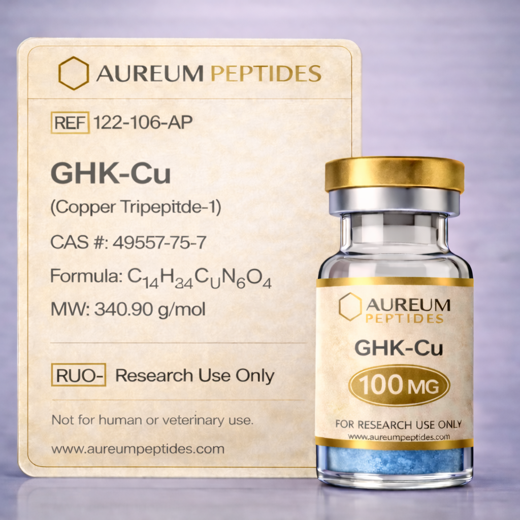
GHK-CU
GHK-Cu, Copper Tripeptide-1, GHK-Copper, Copper Peptide GHK, Glycyl-L-Histidyl-L-Lysine-Copper(II), Cu-GHK, Tripeptide-1 Copper Complex, Gly-His-Lys-Cu
Quality & Transparency
Every batch tested. Every result published. No exceptions.
GHK-Cu is a naturally occurring copper tripeptide complex composed of the tripeptide glycyl-L-histidyl-L-lysine (GHK) bound to a copper(II) ion. First isolated from human plasma in the 1970s, this copper-peptide complex has been extensively studied for its role in extracellular matrix remodeling and cellular signaling processes. The peptide sequence features a high affinity binding site for copper ions through the histidine and terminal amino group, forming a stable coordination complex. GHK-Cu concentrations in biological systems decline with age, prompting research interest in understanding its biochemical mechanisms and cellular effects. In laboratory research, GHK-Cu serves as a valuable tool for investigating copper-dependent signaling pathways, matrix metalloproteinase regulation, and gene expression modulation. Preclinical studies have examined its effects on cellular proliferation, migration, and differentiation across multiple cell types. The compound is classified as a signaling peptide with reported activity in wound healing models, fibroblast function studies, and extracellular matrix synthesis investigations, making it relevant to regenerative biology research applications.
GHK-Cu possesses the amino acid sequence Gly-His-Lys (glycine-histidine-lysine) with molecular formula C₁₄H₂₂N₆O₄Cu and molecular weight of approximately 340 Da when complexed with copper(II). The copper ion coordinates primarily through the histidine imidazole nitrogen, the terminal amino group, and the deprotonated peptide nitrogen, forming a stable square-planar or distorted octahedral geometry depending on pH and additional ligands. This coordination chemistry is critical to its biological activity, as the copper center participates in redox reactions and serves as a cofactor-like element in cellular signaling. The peptide demonstrates stability across physiological pH ranges but is susceptible to proteolytic degradation by peptidases, with copper binding conferring some protection against enzymatic cleavage. GHK-Cu exhibits high affinity for copper ions (log K approximately 16), ensuring stable complex formation under experimental conditions. In aqueous solutions, the compound maintains structural integrity when stored appropriately, though copper dissociation can occur under extreme pH or in the presence of strong chelating agents, factors important for experimental design considerations.
GHK-Cu serves as a versatile research tool in studies investigating copper-dependent biological processes, extracellular matrix dynamics, and cellular signaling mechanisms. Its well-characterized structure and reproducible activity in preclinical models make it valuable for mechanistic investigations across multiple biological systems. The compound has been employed in cell culture studies examining fibroblast activity, keratinocyte behavior, and endothelial cell function, as well as in tissue engineering research exploring scaffold integration and matrix remodeling.
- Gene expression profiling studies examining transcriptional responses to copper-peptide signaling, particularly genes involved in matrix remodeling and antioxidant defense
- Matrix metalloproteinase (MMP) activity assays investigating regulation of MMP-2, MMP-9, and tissue inhibitors of metalloproteinases (TIMPs)
- Cellular migration and proliferation assays in fibroblast, keratinocyte, and endothelial cell models
- Collagen synthesis quantification studies using hydroxyproline assays and immunofluorescence detection of collagen types I and III
- Antioxidant response element activation research examining Nrf2 pathway modulation and downstream target expression
- Angiogenesis model systems including tube formation assays and vascular endothelial growth factor (VEGF) expression studies
- Wound healing research models employing scratch assays, organotypic cultures, and in vivo excisional wound models in rodents
In laboratory research, GHK-Cu has been observed to modulate multiple signaling pathways relevant to tissue remodeling and cellular homeostasis. The compound appears to influence transforming growth factor-beta (TGF-β) superfamily signaling, with in vitro studies indicating effects on Smad pathway activation and downstream target gene expression. This modulation has been associated with altered expression of extracellular matrix components including collagens, fibronectin, and elastin in fibroblast culture systems. Additionally, research suggests GHK-Cu may interact with the epidermal growth factor receptor (EGFR) and related receptor tyrosine kinases, potentially through mechanisms involving copper-mediated redox signaling or direct receptor engagement.
Preclinical studies have demonstrated GHK-Cu effects on matrix metalloproteinase expression and activity, particularly regarding the balance between MMPs and their endogenous inhibitors. In cell culture experiments, the compound has been shown to suppress MMP-1 and MMP-2 expression while upregulating TIMP-1 and TIMP-2, suggesting a role in matrix preservation and remodeling regulation. These effects appear mediated partially through transcription factor modulation, including potential interactions with AP-1 and NF-κB signaling cascades that govern inflammatory and remodeling gene programs.
The copper component of GHK-Cu enables participation in redox-active processes, with research indicating activation of antioxidant response pathways. Studies have reported increased expression of superoxide dismutase (SOD), catalase, and glutathione-related enzymes in cells exposed to GHK-Cu, potentially mediated through Nrf2-ARE (antioxidant response element) pathway activation. This redox activity may contribute to observed effects on cellular stress responses and mitochondrial function in experimental systems. Furthermore, research has identified potential interactions with hypoxia-inducible factor (HIF) pathways and vascular endothelial growth factor (VEGF) signaling, relevant to angiogenesis research models.
Extensive preclinical research has characterized GHK-Cu effects across multiple experimental models. In vitro studies using primary human dermal fibroblasts have demonstrated increased collagen synthesis, enhanced cell proliferation, and altered gene expression profiles favoring anabolic matrix processes. Cell culture experiments have shown that GHK-Cu at micromolar concentrations influences fibroblast migration in scratch assay models and promotes attachment to extracellular matrix substrates. Gene array studies have identified upregulation of genes associated with tissue remodeling, antioxidant defense, and cellular signaling, while simultaneously observing downregulation of inflammatory mediators and matrix-degrading enzymes. Research using keratinocyte cultures has indicated effects on differentiation markers and proliferation rates relevant to epithelialization processes.
In vivo rodent models have provided data on wound healing kinetics, with studies reporting accelerated closure rates, enhanced granulation tissue formation, and improved histological characteristics in GHK-Cu-treated wounds compared to vehicle controls. These observations have been accompanied by biochemical measurements showing increased hydroxyproline content (indicating collagen deposition), altered cytokine profiles, and enhanced angiogenesis markers. Immunohistochemical analyses have revealed increased expression of collagen types I and III, as well as markers of neovascularization including CD31 and VEGF. Time-course studies in excisional wound models have documented effects on inflammatory cell infiltration, myofibroblast presence, and epithelial migration patterns.
Additional preclinical research has examined GHK-Cu in models of oxidative stress, with cell culture studies demonstrating protective effects against hydrogen peroxide and other oxidative insults. These observations correlate with measured increases in antioxidant enzyme activities and reduced markers of oxidative damage. Endothelial cell research has documented effects on tube formation assays, migration responses, and VEGF expression, supporting investigation of angiogenic mechanisms. Proteomic and transcriptomic analyses have identified broad effects on cellular signaling networks, extracellular matrix composition, and metabolic pathways, providing molecular-level context for observed phenotypic changes in experimental systems.
GHK-Cu is supplied as a lyophilized powder, a form that provides optimal stability during storage and shipping while facilitating precise reconstitution for experimental applications. Each production batch undergoes comprehensive analytical verification to ensure consistency and quality for research use. High-performance liquid chromatography (HPLC) analysis confirms purity specifications, typically ≥98%, by separating and quantifying the peptide-copper complex and potential impurities. Mass spectrometry provides molecular weight confirmation and structural identity verification, ensuring the correct peptide sequence and copper coordination. Endotoxin testing using Limulus Amebocyte Lysate (LAL) assay confirms levels below specified thresholds, critical for cell culture applications where endotoxin contamination could confound experimental results. A Certificate of Analysis (COA) accompanies each product lot, documenting these analytical results and providing traceability for laboratory records. These quality control measures support reproducibility and reliability in research workflows, enabling investigators to implement consistent experimental protocols across studies.
- 1Pickart L et al., Journal of Biological Chemistry, 1980 255(3):1064-1069. PubMed
- 2Pickart L and Thaler MM, Nature, 1973 243(5408):85-87. PubMed
- 3Choi HR et al., Archives of Pharmacal Research, 2012 35(2):175-182. PubMed
- 4Park JR et al., Journal of Dermatological Science, 2012 66(2):175-178. PubMed
- 5Pollard JD et al., Journal of Peptide Science, 2011 17(11):770-778. PubMed
- 6Canapp SO et al., International Wound Journal, 2003 1(1):25-32. PubMed
- 7Amin S et al., Skin Pharmacology and Physiology, 2004 17(5):207-213. PubMed
- 8Kang YA et al., Biotechnology Letters, 2009 31(2):209-213. PubMed
- 9Hostynek JJ et al., Food and Chemical Toxicology, 2011 49(8):1887-1893. PubMed
- 10Campo GM et al., Biochemical Pharmacology, 2008 75(2):594-611. PubMed
- 11Pyo SM et al., Cancer Letters, 2011 308(1):79-90. PubMed
- 12Wang X et al., International Journal of Molecular Sciences, 2017 18(12):2674. PubMed
Advancing Research, One Peptide at a Time
Premium quality. Rigorous testing. Trusted by researchers worldwide.

Third-party testing data will be displayed here once available.
Temperature
Lyophilized: -20°C
Reconstituted: 2-8°C (30 days)
Shelf Life
24 months lyophilized
30 days reconstituted
Handling
Avoid freeze-thaw cycles.
Use bacteriostatic water for reconstitution.
Frequently Asked Questions
Stay at the Forefront
Join our research community. Get early access to new peptides, exclusive member pricing, and curated literature reviews delivered to your inbox.
Welcome to the Circle ✦
Check your inbox for your 10% welcome discount
Related Peptides
Explore complementary research compounds